
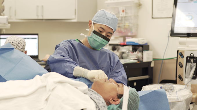
The Definitive Treatment
DMEK & DSEK Surgery
Endothelial keratoplasty replaces only the diseased Descemet's membrane and endothelial cells — the two innermost layers of the cornea. The result: a definitive, lasting restoration of corneal clarity with minimal disruption to ocular anatomy, through a small incision.
5000+
Ocular Surgeries
12yrs+
Surgical Experience
45 min
Procedure Time
— Choosing Your Procedure
DMEK or DSEK —
Which is Right For You?
DMEK
Descemet's Membrane Endothelial Keratoplasty
The most selective transplant in ophthalmology. Only Descemet's membrane and the endothelial monolayer are transplanted — a self-scrolling ribbon of tissue just 10–15μm thick — leaving the recipient stroma entirely undisturbed. The thinner graft means no interface scatter, yielding superior visual acuity.
10-20μm
Graft Thickness
<3mm
Incision size
20/20-30 vision
Typical Outcome
1-3 month
Recovery Time
No stromal tissue transplanted — no interface haze
Superior visual acuity compared to DSEK
Fastest visual recovery of all keratoplasty techniques
Near-zero endothelial rejection risk
Minimal disruption to ocular anatomy — changes barely visible
Both procedures are endothelial keratoplasty — both replace only the diseased inner layer and both eliminate the risk of stromal rejection. The choice between them depends on your corneal anatomy, ocular history, and the specific judgment of a corneal surgeon.
DSEK
Descemet's Stripping Endothelial Keratoplasty
DSEK transplants a thin disc of donor tissue including a thin layer of posterior stroma with Descemet's membrane and endothelium attached. Less technically demanding than DMEK, and preferred in anatomically complex eyes — shallow anterior chambers, prior vitrectomy, iris defects, or challenging pupil dynamics.
50–120μm
Graft Thickness
3-5mm
Incision Size
20/20-30 vision
Typical Outcome
1-3 month
Recovery Time
Preferred for anatomically complex or previously operated eyes
Suitable for prior vitrectomy, iris abnormalities, shallow AC
Very low rejection risk — endothelial graft only
No sutures required; air bubble tamponade
Appropriate when DMEK tissue handling is not feasible
— Surgical Process
The DSEK/DMEK Transplant Journey,
Step by Step
Evaluation
Candidacy Assessment
Preparation
Tissue Procurement & Preparation
Surgery
The Procedure
Recovery
Post-Operative Care
Clinical evaluation noting endothelial cell loss and guttae and/or edema. Anterior segment OCT maps anterior chamber depth — essential for DMEK eligibility. Dr. Hu reviews all data to determine whether DMEK or DSEK is the optimal approach for your eye.
RECOVERY & FOLLOW-UP
Patients rest face-up for several hours after surgery to maintain air tamponade. Vision begins improving within days as the graft adheres and pumps residual oedema from the stroma. A small percentage of patients (~10%) require a simple in-office "rebubbling" procedure if partial graft detachment occurs. Topical steroids are tapered over six months. Dr. Hu performs all follow-up examinations personally.
THE PROCEDURE
Under topical anaesthesia, a small peripheral incision (<3mm for DMEK) allows introduction of a glass injector. The diseased Descemet's membrane is scored and stripped from the recipient cornea. The donor scroll is injected, orientated using ink markings, and unrolled inside the anterior chamber using careful fluid and air manoeuvres. An air bubble is injected to tamponade the graft against the recipient stroma.
PRE-OPERATIVE OPTIMISATION
Donor tissue is sourced from EBAA-accredited eye banks with full serological screening and pre-cut endothelial cell density verification. For DMEK, the Descemet's membrane is carefully peeled from the donor stroma and prepared as a thin scroll — a technically demanding process performed under operating microscope guidance.
Endothelial Keratoplasty (DSAEK & DMEK) Surgery
Endothelial Keratoplasty (EK) is a cornea transplant technique that can restore lost vision when the endothelium, the innermost layer of cells of the cornea, are no longer functioning adequately.
There are a two variants of EK surgery:
Descemets’ Stripping Automated Endothelial Keratoplasty (DSAEK)
Descemets’ Membrane Endothelial Keratosplaty (DMEK)
Medical conditions including Fuchs’ dystrophy, bullous keratopathy, iridocorneal endothelial (ICE) syndrome, or other endothelial disorders may cause blurry or cloudy vision and glare. Vision is typically worse in the morning and clears up throughout the day. An EK procedure selectively replaces only the diseased layer of the cornea, leaving healthy areas intact.
There are many advantages of an EK surgery over a full-thickness Penetrating Keratoplasty surgery, including:
faster recovery time
lower risk, and less invasive surgery
fewer sutures in the eye, and a lower chance of an infection
better long-term quality of vision
lower long-term risk of graft rejection
What is the difference between a DSAEK surgery and DMEK surgery?
A Descemets’ membrane endothelial keratoplasty (DMEK) is very similar to DSAEK, except that the implanted donor tissue does not include any stromal tissue. A DMEK is an exciting option to treat decreased vision and swollen cloud corneas and it is a pure replacement of endothelium. DMEK is the most anatomical repair of the three generations of corneal transplantation with just one cell layer and a thin membrane, all of which are only 15 to 20 microns thick.
DMEK Procedure
15-20 microns of endothelial cell layer and Descemets’ membrane transplanted into the eye
Minimal disruption of ocular anatomy
lowest lifetime risk of graft rejection
Patients have a very high quality of vision afterwards, many achieving vision of 20/25 or better
DSAEK Procedure
60-120 microns of corneal stroma, along with endothelial cell layer and Descemets’ membrane transplanted into the eye
Can be performed in patients with complex ocular anatomy (prior corneal transplant, history of glaucoma, prior trauma)
Easier post-operative recovery experience
Patients have a very good quality of vision afterwards, many achieving vision of 20/30 or better
What To Expect During a DSAEK or a DMEK Procedure
During both DSAEK and DMEK surgery, the diseased innermost layer of the cornea is removed and the thin layer from a healthy donor cornea is put in its place. The transplant is then held in place by only an air bubble. Patients will be required to lay flat on their backs with their faces directed upwards immediately after surgery to float the bubble into place. For patients who have had DSAEK, the patient will have to lay flat on their back for 24-36 hours after surgery while the air bubble dissipates. For patients who have had DMEK surgery, the air bubble is mixed with SF6 gas, and the patient will have to lay flat on their back for 4-5 days after surgery.
Your doctor will discuss with you the options that will better suit your needs.
DMEK vs DSEK surgery
What is the advantage of DMEK surgery (compared to DSEK surgery)?
DMEK has been shown to offer patients the best chance to see 20/25 and resume their daily activities quickly. There is also a slightly lower long-term risk of graft rejection with DMEK compared to DSEK.
What is the disadvantage of DMEK surgery (compared to DSEK surgery)?
A patient has to lay flat for a longer time after DMEK surgery, compared to DSEK surgery, because the thinner graft does not stick/adhere to the host cornea quite as well. A longer period of time is needed to ensure that the graft adheres well. The chance of a graft detachment is also much higher.
Your doctor will discuss with you the options that will better suit your needs.
When is DSEK preferred over DMEK?
Usually DSEK is preferred over DMEK surgery when a patient cannot lay flat long enough for DMEK, or has issues laying flat in general. Patients with neck, spine issues or back pain in general may benefit from DSEK surgery. Complicated EK surgery generally favors DSEK. Patients with other ocular conditions (a history of a prior corneal transplant, prior eye surgery/trauma, a history of glaucoma) typically benefit more from DSEK surgery

An eye exam is the best way to screen for, and evaluate any eye conditions affecting your health, comfort, and quality of vision.